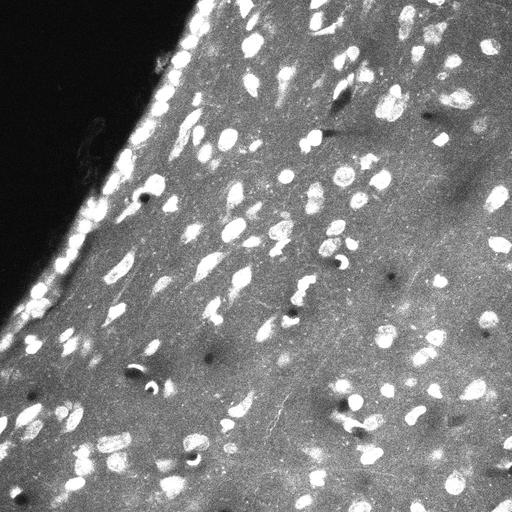
In this study, we developed an electron microscopy method, called CryoChem, to optimally preserve the morphology of genetically labeled tissues. This method also allows us to perform 3D correlated light and electron microscopy (CLEM) in the well-preserved tissue. This dataset contains the DRAQ5 confocal volume of the CLEM experiment in a mouse brain expressing tdTomato in corticotropin releasing factor-expressing neurons.
Brief description of sample preparation
After cryofixation by high-pressure freezing and freeze-substitution, cryofixedmouse brain slices were rehydrated gradually. Rehydrated samples were then imaged with confocal microscopy to capture the DRAQ5 and tdTomato signals. Next, the samples were stained using a high-contrast en bloc staining protocol. Then the samples were dehydrated for resin infiltration and embedding, followed by imaging with X-ray microscopy and then SBEM.
Confocal Microscopy
After freeze-substitution and rehydration, the mouse brain slice was placed in ice-cold 0.15 M sodium cacodylate for imaging. Confocal volumes of DRAQ5 and tdTomato signals were collected on an Olympus FluoView 1000 confocal microscope with a 20X air and 60X water objectives using 561 nm and 633 nm excitation.
| Spatial Axis | Image Size | Pixel Size |
|---|---|---|
| X | 1024px | —— |
| Y | 1024px | —— |
| Z | 73px | —— |