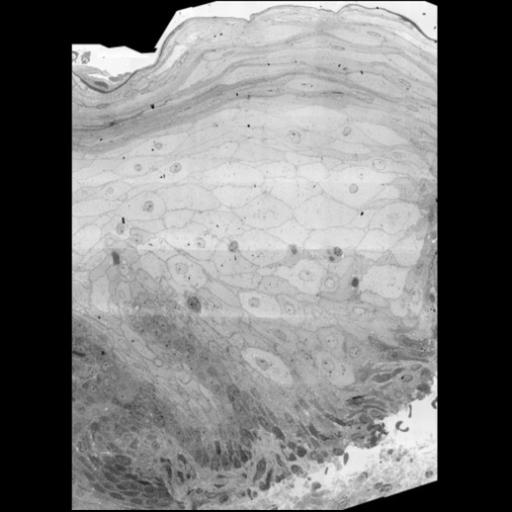
High-resolution montage of a thin section of Healthy human skin, including part of the dermis and all epidermal layers. This image is a montage of 32 images and a series of 10 datasets as published: http://www.jidonline.org/article/S0022-202X(15)37322-X/fulltext http://www.nanotomy.org/OA/Sokol2015JID/index.html
Two mm skin were fixed in 2% glutaraldehyde in 0.1 M sodium cacodylate buffer pH 7.4 washed in 0.1 M sodium cacodylate buffer and postfixed in 1% osmiumtetroxide and 1.5% potassiumferrocyanide. Samples were dehydrated, embedded in epon, and sectioned. Ultrathin sections (70 nm) were positioned on single slot holders A600 Nickel supported by Formvar and contrasted with 2% uranylacetate (methanol) followed by Reynolds lead citrate. Acquisition was performed using a Zeiss supra 55 EM (Oberkochen, Germany) with ATLAS software developed by Fibics (Ottawa, Ontario, Canada). Samples were recorded at 2.5 nm pixel size. Scans were stitched, and raw data sets were rendered as HTML files using ATLAS VE viewer software. data sets are orientated such that the epidermis/superficial layers are facing up.
| Spatial Axis | Image Size | Pixel Size |
|---|---|---|
| X | 234px | 93.696µm |
| Y | 326px | 130.560µm |
| Z | 326px | 130.560µm |