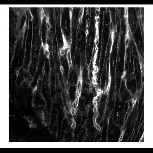
Images are acquired from fixed and living heart tissue using fiber-optics and laser-scanning confocal microscopy, respectively. Three sets of images denoted as CCM, FCMtopical, FCMcarrier were stored for subsequent analyses.
CCM images were from fixed rodent tissue preparations acquired using established methods (doi: 10.1161/circimaging.112.000121). In short, rodent hearts were isolated and Langendorff-perfused with 4% paraformaldehyde. Tissue from the atrial working myocardium, sinoatrial node, and atrioventricular node were dissected from the hearts and fluorescently labeled with wheat germ agglutinin conjugated to CF488A (29022-1; Biotium, Hayward, CA; 1:25). Fluorescently labeled tissue preparations were imaged using a conventional laser-scanning confocal microscope (Zeiss LSM5 Duo; Zeiss, Jena, Germany) with a 40x oil immersion lens (NA 1.3). The fluorescent signal was captured using an Argon/2 ion laser (488 nm excitation) and a bandpass filter (505 to 555 nm emission). Three-dimensional image stacks were acquired at a spatial resolution (xyz dimensions), field of view (xy) and a z-scan range of 0.2x0.2x0.2 μm, 204.8x204.8 μm and 50 μm, respectively. Example cross-sections through the image stacks were stored as CCM images.
| Spatial Axis | Image Size | Pixel Size |
|---|---|---|
| X | —— | 204.8µm |
| Y | —— | 204.8µm |
| Z | —— | 50µm |