Alternate header for print version


Licensing
Public Domain: This image is in the public domain and thus free of any copyright restrictions. However, as is the norm in scientific publishing and as a matter of courtesy, any user should credit the content provider for any public or private use of this image whenever possible. Learn more
tweet  
*CIL – Cell Image Library accession number. Please use this to reference an image.

CIL:4665*  Cite 
Description

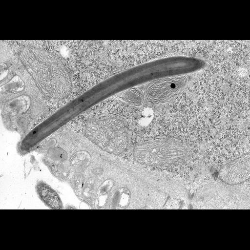
A high resolution image of the cortex of Didinium nasutum. Embedded within the fibrous layer are many extrusive organelles, called mucocysts, involved in cyst formation. In this view there is also a longitudinal section of a more prominent and long curved extrusome (called a cyrtocyst by Wessenberg and Antipa, 1968) in the proter of a dividing cell. The cyrtocyst is of unknown function. Extrusomes of Didinium typically have a bundle of myelin-like membrane material enclosed by their surrounding membrane. This may be stored membrane needed for extrusome discharge. TEM taken on 5/20/69 by R. Allen with a Philips 300 operating at 60kV. Neg. 14,800X. The raw film was scanned with a Nikon Coolscan 9000ED. Standard glutaraldehyde fixation followed by osmium tetroxide, dehydrated in alcohol and embedded in an epoxy resin. Microtome sections prepared at approximately 75nm thickness. Additional information available at (http://www5.pbrc.hawaii.edu/allen/).

Biological Sources
NCBI Organism Classification
Didinium nasutum
Cell Type
eukaryotic cell
Eukaryotic Protist
Ciliated Protist
Cell Line
Carolina Biological Supply Company, NC, U.S.A.
Cellular Component
cell cortex
extrusive organelles
mucocyst
cyrtocyst
Biological Context
Biological Process
cortical organization
cyst formation
unknown function
Attribution
Names
Richard Allen
Citation
Digital Object Identifier (DOI)
doi:10.7295/W9CIL4665
Archival Resource Key (ARK)
ark:/b7295/w9cil4665
Grouping This image is part of a group.
Imaging
Image Type
transmission electron microscopy (TEM)
illumination by electrons
Image Mode
detection of electrons
film
Parameters Imaged
electron density
Source of Contrast
stain with broad specificity
Visualization Methods
stain with broad specificity
osmium tetroxide
uranyl salt
lead salt
Processing History
film
unprocessed raw data
Data Qualifiers
raw, unprocessed data
suitable for spatial measurements
Dimensions
Spatial Axis Image Size Pixel Size
X 5516px 1nm
Y 3745px 1nm