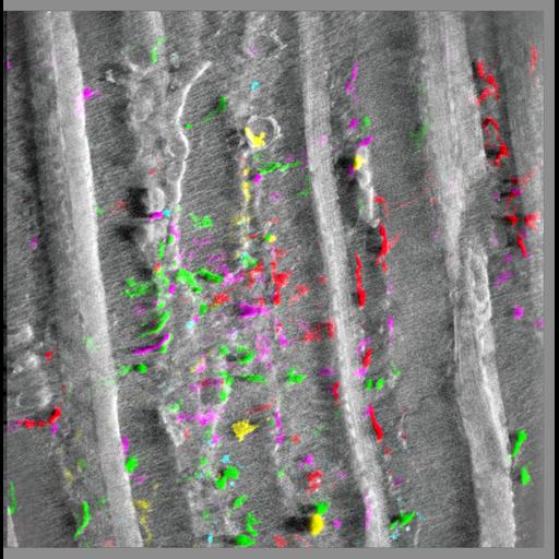
The picture represents a 3D-reconstruction (using Imaris software) of confocal/multiphoton images collected through the thickness (100 micrometers) of the live skin from a bone-marrow transplanted mouse. Cells marked by five fluorescent proteins: Cerulean (cyan), EGFP (green), Venus (yellow), tdTomato (purple), mCherry (red) appear interspersed along reticulin and collagen fibers (second harmonic generation-SHG, white) at 4 months post-transplant. See also Malide et al. (2012) Dynamic clonal analysis of murine hematopoietic stem and progenitor cells marked by 5 fluorescent proteins using confocal and multiphoton microscopy. Blood. Dec 20;120(26):e105-16. This image is grouped with two others obtained in a similar fashion.
Imaging was performed using a Leica TCS SP5-AOBS 5-channel confocal/two-photon system equipped with multiline Argon diode 561 nm, HeNe 594 nm and HeNe 633 nm as well as TiSaph two-photon laser. Images were recorded with a 25x 0.95 NA water dipping objective lens, and 5 micrometer z-spacing and processed using Imaris software (Bitplane). See also: Malide D, Metais J-Y, and Dunbar CE. (2012). Dynamic clonal analysis of murine hematopoietic stem and progenitor cells marked by 5 fluorescent proteins using confocal and multiphoton microscopy. Blood. Dec 20;120(26):e105-16.
| Spatial Axis | Image Size | Pixel Size |
|---|---|---|
| X | 939px | —— |
| Y | 952px | —— |