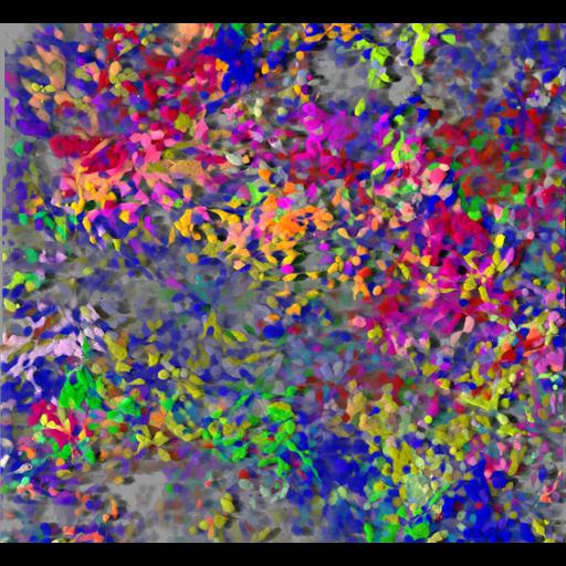
The image shows a 3D-rendering (Imaris software) of a live confluent culture of NIH-3T3 cells obtained using confocal microscopy. The cells were co-transduced with 5 fluorescent proteins and with Lentiviral Gene Ontology (LeGO) vectors expressing Cerulean (blue), EGFP (green), Venus (yellow), tdTomato (magenta) or mCherry (red) fluorescent proteins to provide combinatorial colors for progeny tracking. Groups of nearby cells of the same color descended from same stem cells. This image is grouped with two others obtained using a similar imaging strategy. See also Malide et al. (2012) Dynamic clonal analysis of murine hematopoietic stem and progenitor cells marked by 5 fluorescent proteins using confocal and multiphoton microscopy. Blood. Dec 20;120(26):e105-16. An image obtained in the same manner that obtained an Honorable Mention in the 2011 Olympus BioScapes Digital Imaging Competition may be viewed as CIL:41631.
Imaging was performed using a Leica TCS SP5-AOBS 5-channel confocal system equipped with multiline Argon diode 561 nm, HeNe 594 nm and HeNe 633 nm. Images were recorded with a 40x 1.4 NA oil immersion objective lens, and 5 micrometer z-spacing and processed using Imaris software (Bitplane). See also: Malide D, Metais J-Y, and Dunbar CE. (2012). Dynamic clonal analysis of murine hematopoietic stem and progenitor cells marked by 5 fluorescent proteins using confocal and multiphoton microscopy. Blood. Dec 20;120(26):e105-16.
| Spatial Axis | Image Size | Pixel Size |
|---|---|---|
| X | 1104px | —— |
| Y | 1014px | —— |