Alternate header for print version


Licensing
Attribution Only: This image is licensed under a Creative Commons Attribution License. View License Deed | View Legal Code
tweet  
*CIL – Cell Image Library accession number. Please use this to reference an image.

CIL:43601*  Cite 
Description

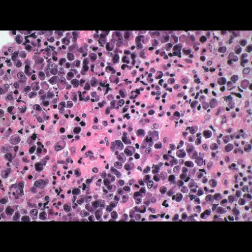
Shown is a hematoxylin and eosin stained section through a plasmablastic lymphoma of the oral cavity in an HIV-positive adult male. There is a diffuse proliferation of large neoplastic cells most of which resemble B immunoblasts, plasmablasts or atypical plasma cells.

Technical Details

Stained sections were recorded with a CCD camera mounted on a Nikon Eclipse 80i light microscope using a 40x objective lens.

Biological Sources
NCBI Organism Classification
Homo sapiens
Cell Type
lymphocyte
Biological Context
Biological Process
immune response to tumor cell
Human Disease
Human immunodeficiency virus infectious disease
Attribution
Names
Gabriel Caponetti
Citation
Digital Object Identifier (DOI)
doi:10.7295/W9CIL43601
Archival Resource Key (ARK)
ark:/b7295/w9cil43601
Imaging
Image Type
recorded image
Image Mode
bright-field microscopy
Parameters Imaged
absorption of illumination
Source of Contrast
differences in adsorption or binding of stain
Visualization Methods
hematoxylin
eosin
Sample Preparation
Methods
formaldehyde fixed tissue
tissue in wax embedment
Relation To Intact Cell
sectioned tissue
Dimensions
Spatial Axis Image Size Pixel Size
X 2296px ——
Y 1744px ——