Alternate header for print version


Licensing
Public Domain: This image is in the public domain and thus free of any copyright restrictions. However, as is the norm in scientific publishing and as a matter of courtesy, any user should credit the content provider for any public or private use of this image whenever possible. Learn more
tweet  
*CIL – Cell Image Library accession number. Please use this to reference an image.

CIL:43552*  Cite 
Description

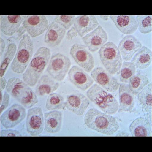
Light micrograph of onion (Allium cepa) root tip cells stained with acetocarmine to show nuclei and chromosomes. The field includes cells in interphase, prophase, metaphase, and late telophase.

Technical Details

Acetocarmine stained root tips were viewed with a bright field light microscope using a 40x 0.65 N.A. objective lens, and recorded on film.

Biological Sources
NCBI Organism Classification
Allium cepa
Cell Type
root tip
Cellular Component
nucleus
condensed chromosome
Biological Context
Biological Process
mitosis
Attribution
Names
Shoba Shanti
Citation
Digital Object Identifier (DOI)
doi:10.7295/W9CIL43552
Archival Resource Key (ARK)
ark:/b7295/w9cil43552
Imaging
Image Type
film
Image Mode
modes of light microscopy
Parameters Imaged
compartmentalization of stain or label
Source of Contrast
differences in adsorption or binding of stain
Visualization Methods
acetocarmine
Data Qualifiers
processed data
suitable for spatial measurements
Sample Preparation
Methods
chemically fixed tissue
Relation To Intact Cell
whole mounted tissue
Dimensions
Spatial Axis Image Size Pixel Size
X 1280px ——
Y 1024px ——