Alternate header for print version


Licensing
Attribution Non-Commercial; No Derivatives:This image is licensed under a Creative Commons Attribution, Non-Commercial, No Derivatives License. View License Deed | View Legal Code
tweet  
*CIL – Cell Image Library accession number. Please use this to reference an image.

CIL:42508*  Cite 
Description

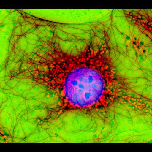
Digital multi-color immuno fluorescence microscopy and phase contrast image of fibroblast cells in culture stained with various antibodies. The Phase contrast (green) image was overlayed over the microtubule (red) and nucleus (Blue) image. Honorable Mention, 2007 Olympus BioScapes Digital Imaging Competition

Technical Details

Biological Sources
Cell Type
fibroblast
Cellular Component
microtubule cytoskeleton
nucleus
Biological Context
Biological Process
cytoskeleton organization
Attribution
Names
Jan Schmoranzer
2007 Olympus BioScapes Digital Imaging Competition
Link
2007 Olympus BioScapes Digital Imaging Compe...
OTHER
New York, NY, USA
Citation
Digital Object Identifier (DOI)
doi:10.7295/W9CIL42508
Archival Resource Key (ARK)
ark:/b7295/w9cil42508
Sample Preparation
Methods
chemically fixed tissue
permeabilized tissue
Relation To Intact Cell
dispersed cells in vitro
Dimensions
Spatial Axis Image Size Pixel Size
X 2000px ——
Y 1763px ——