Alternate header for print version


Licensing
Attribution Non-Commercial Share Alike:This image is licensed under a Creative Commons Attribution, Non-Commercial Share Alike License. View License Deed | View Legal Code
tweet  
*CIL – Cell Image Library accession number. Please use this to reference an image.

CIL:42256*  Cite 
Description

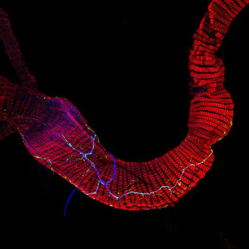
Confocal micrograph of the posterior portion of the digestive tract of an adult Drosophila fly. The circular visceral muscles are labelled in red with phalloidin staining of actin. The main nerves are stained in blue (with 22c10, a MAP1B antibody), and an insulin-like peptide (Ilp7), released by a subset of gut-innervating neurons, is labelled in green.

Biological Sources
NCBI Organism Classification
Drosophila melanogaster
Cell Type
muscle cell
neuron
Cellular Component
actin filament
ILP7
neuron projection
MAP1B
Biological Context
Biological Process
insulin-like peptide release
Attribution
Names
Irene Miguel-Aliaga
OTHER
Department of Zoology, University of Cambridge. Downing Street, Cambridge, CB2 3EJ, United Kingdom
Citation
Digital Object Identifier (DOI)
doi:10.7295/W9CIL42256
Archival Resource Key (ARK)
ark:/b7295/w9cil42256
Grouping This image is part of a group.
Imaging
Image Type
recorded image
Image Mode
confocal microscopy
Parameters Imaged
fluorescence emission
Source of Contrast
distribution of epitope
Visualization Methods
primary antibody plus labeled secondary antibody
phalloidin
Sample Preparation
Methods
formaldehyde fixed tissue
Relation To Intact Cell
whole mounted tissue
Dimensions
Spatial Axis Image Size Pixel Size
X 1024px ——
Y 1024px ——