Alternate header for print version


Licensing
Attribution Non-Commercial; No Derivatives:This image is licensed under a Creative Commons Attribution, Non-Commercial, No Derivatives License. View License Deed | View Legal Code
tweet  
*CIL – Cell Image Library accession number. Please use this to reference an image.

CIL:41631*  Cite 
Description

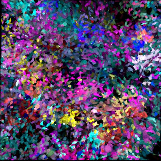
Confocal micrograph of NIH-3T3 cells co-transduced with 5 fluorescent proteins. The cells were marked by co-transduction with Lentiviral Gene Ontology (LeGO) vectors expressing Cerulean (cyan), EGFP (green), Venus (yellow), tdTomato (magenta) or mCherry (red) fluorescent proteins to provide combinatorial colors for cell lineage tracing. These are cytosolic-untargeted FPs (just to mark the cells). The image was collected on live cells with a Leica SP5 confocal system. Honorable Mention, 2011 Olympus BioScapes Digital Imaging Competition®.

Technical Details

Biological Sources
NCBI Organism Classification
Mus musculus
Cell Type
fibroblast
Cell Line
NIH/3T3
Cellular Component
cytoplasm
Attribution
Names
Daniela Malide
2011 Olympus BioScapes Digital Imaging Competition®
Link
2011 Olympus BioScapes Digital Imaging Compet...
OTHER
National Institutes of Health, Bethesda, MD, USA
Citation
Digital Object Identifier (DOI)
doi:10.7295/W9CIL41631
Archival Resource Key (ARK)
ark:/b7295/w9cil41631
Imaging
Image Type
recorded image
Image Mode
confocal microscopy
Parameters Imaged
fluorescence emission
Source of Contrast
compartmentalization of stain or label
Visualization Methods
CeruleanFP
EGFP
VenusFP
tdTomatoFP
mCherryFP
Sample Preparation
Methods
living tissue
Relation To Intact Cell
dispersed cells in vitro
Dimensions
Spatial Axis Image Size Pixel Size
X 1976px ——
Y 1966px ——