Licensing

Attribution_by_nc_nd
Attribution Non-Commercial; No Derivatives:This image is licensed under a Creative Commons Attribution, Non-Commercial, No Derivatives License. View License Deed | View Legal Code
Comments: Powered by Pnyxe

Description

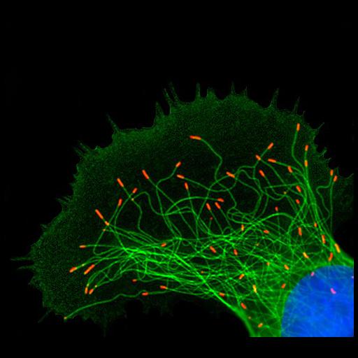
Fluorescent micrograph of a Xenopus melanophore, showing microtubules (green), microtubule plus ends (red) and nucleus (blue). Honorable Mention, 2011 Olympus BioScapes Digital Imaging Competition®.

Technical Details

Biological Sources

NCBI Organism Classification
Xenopus
(Clawed frogs)
Cell Type
melanocyte
Cellular Component
microtubule cytoskeleton
nucleus

Attribution

Names
Alexis J. Lomakin
2011 Olympus BioScapes Digital Imaging Competition®
Link
2011 Olympus BioScapes Digital Imagin...
Other
Howard Hughes Medical Institute, Carnegie Institution of Washington, Baltimore, MD, USA

Imaging

Image Type
recorded image
Imaging Mode
fluorescence microscopy
Parameters Imaged
fluorescence emission

Sample Preparation

Relation To Intact Cell
dispersed cells in vitro

Dimensions

Spatial Axis Image Size Pixel Size
X 500px ——
Y 452px ——
*CIL – Cell Image Library accession number. Please use this to reference an image.