Alternate header for print version


Licensing
Public Domain: This image is in the public domain and thus free of any copyright restrictions. However, as is the norm in scientific publishing and as a matter of courtesy, any user should credit the content provider for any public or private use of this image whenever possible. Learn more
tweet  
*CIL – Cell Image Library accession number. Please use this to reference an image.

CIL:41604*  Cite 
Description

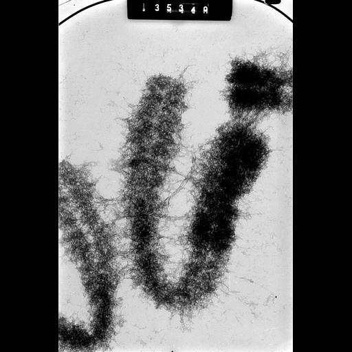
High Voltage Transmission EM of whole mitotic metaphase chromosomes from Chinese hamster ovary cells. This image was recorded at a specimen tilt of 58 degrees and is grouped with one taken at 48 degrees, creating a stereo pair giving an oblique 3D view of the chromosomes.

Technical Details

CHO cells arrested at metaphase with colcemid were treated with 67% D2O, in 0.07M phosphate buffer and squashed in 50% acetic acid. Preparations were transferred to EM grids and examined with the Wisconsin HVEM operated at 1MeV. See also: H. Ris 1981 Stereoscopic electron microscopy of chromosomes. Meth Cell Biol 22:77-96 H. Ris 1978 Preparation of chromatin and chromosomes for electron microscopy. Meth Cell Biol 18:220-246.

Biological Sources
NCBI Organism Classification
Cricetulus griseus
Cell Type
ovary
epithelial cell
Cell Line
CHO
Cellular Component
nuclear chromosome
Biological Context
Biological Process
chromosome organization
mitotic metaphase
mitosis
Attribution
Names
Hans Ris
Citation
Digital Object Identifier (DOI)
doi:10.7295/W9CIL41604
Archival Resource Key (ARK)
ark:/b7295/w9cil41604
Grouping This image is part of a group.
Imaging
Image Type
film
Image Mode
Parameters Imaged
electron density
Source of Contrast
differences in adsorption or binding of stain
Visualization Methods
uranyl salt
Data Qualifiers
raw, unprocessed data
suitable for spatial measurements
Sample Preparation
Methods
chemically fixed tissue
Relation To Intact Cell
squash preparation
Dimensions
Spatial Axis Image Size Pixel Size
X 2821px 2nm
Y 4234px 2nm