Alternate header for print version


Licensing
Attribution Non-Commercial; No Derivatives:This image is licensed under a Creative Commons Attribution, Non-Commercial, No Derivatives License. View License Deed | View Legal Code
tweet  
*CIL – Cell Image Library accession number. Please use this to reference an image.

CIL:41553*  Cite 
Description

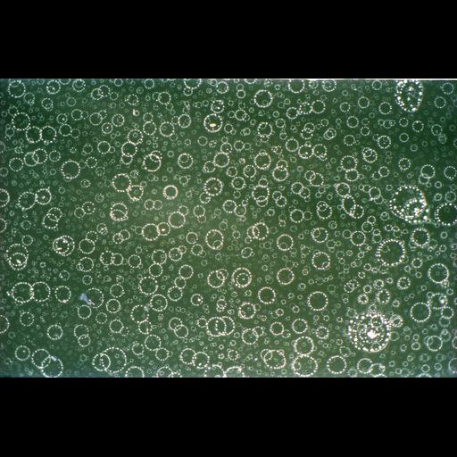
Phase contrast image of serum arrested mouse L-1210 cells engaged in spontaneous apoptosis (programmed cell death) after nutrient depletion and acid hydrolysis. The image was taken at 40X, scanned, and then enlarged. Honorable Mention, 2011 Olympus BioScapes Digital Imaging Competition®.

Technical Details

Biological Sources
NCBI Organism Classification
Mus musculus
Cell Type
lymphocytic leukemia
Cell Line
L1210
Cellular Component
cell surface
Biological Context
Biological Process
induction of apoptosis
cellular response to nutrient levels
acid hydolysis
Attribution
Names
Frank Abernathy
2011 Olympus BioScapes Digital Imaging Competition®
Link
2011 Olympus BioScapes Digital Imaging Compet...
OTHER
Jamestown, OH, USA
Citation
Digital Object Identifier (DOI)
doi:10.7295/W9CIL41553
Archival Resource Key (ARK)
ark:/b7295/w9cil41553
Imaging
Image Type
recorded image
Image Mode
phase contrast microscopy
Parameters Imaged
elastic scattering of photons
Source of Contrast
differences in amount of elastic light scattering
Visualization Methods
visualization of contiguous regions
Processing History
scanned
enlarged
Sample Preparation
Relation To Intact Cell
dispersed cells in vitro
Dimensions
Spatial Axis Image Size Pixel Size
X 6753px ——
Y 4427px ——