Alternate header for print version


Licensing
Public Domain: This image is in the public domain and thus free of any copyright restrictions. However, as is the norm in scientific publishing and as a matter of courtesy, any user should credit the content provider for any public or private use of this image whenever possible. Learn more
tweet  
*CIL – Cell Image Library accession number. Please use this to reference an image.

CIL:41482*  Cite 
Description

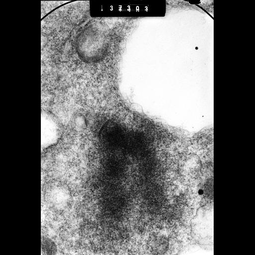
High voltage (1MeV) transmission electron microscopy image of a 0.5 micrometer section through a Chinese hamster ovary cell at metaphase showing a chromatid pair, with kinetochores at the centromere. The image was taken with a specimen tilt of 55 degrees and is grouped with one at 40 degrees. The pair provides an oblique stereo view of the chromosome.

Technical Details

CHO cells at metaphase were exposed to 0.07M KCl, fixed with paraformaldehyde followed by OsO4, embedded in epoxy resin and 0.5 micrometer 'thick' sections stained with uranly acetate for HVEM observation. See also: H. Ris 1981 Stereoscopic electron microscopy of chromosomes. Meth Cell Biol 22:77-96 H. Ris 1978 Preparation of chromatin and chromosomes for electron microscopy. Meth Cell Biol 18:220-246.

Biological Sources
NCBI Organism Classification
Cricetulus griseus
Cell Type
epithelial cell
Cell Line
CHO
Cellular Component
nuclear chromosome
kinetochore
Biological Context
Biological Process
chromosome organization
mitosis
mitotic metaphase
Attribution
Names
Hans Ris
Citation
Digital Object Identifier (DOI)
doi:10.7295/W9CIL41482
Archival Resource Key (ARK)
ark:/b7295/w9cil41482
Grouping This image is part of a group.
Imaging
Image Type
film
Image Mode
Parameters Imaged
electron density
Source of Contrast
differences in adsorption or binding of stain
Visualization Methods
uranyl salt
osmium tetroxide
Data Qualifiers
raw, unprocessed data
suitable for spatial measurements
Sample Preparation
Methods
formaldehyde fixed tissue
tissue in plastic embedment
Relation To Intact Cell
microtome-sectioned tissue
Dimensions
Spatial Axis Image Size Pixel Size
X 2876px 1nm
Y 4238px 1nm