Alternate header for print version


Licensing
Attribution Non-Commercial; No Derivatives:This image is licensed under a Creative Commons Attribution, Non-Commercial, No Derivatives License. View License Deed | View Legal Code
tweet  
*CIL – Cell Image Library accession number. Please use this to reference an image.

CIL:41465*  Cite 
Description

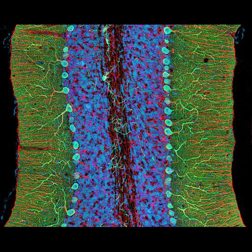
Photomicrograph of a midsaggital section showing the different components of the rat cerebellum, including Purkinje neurons in green, glia (non-neuronal cells) in red, and cell nuclei in blue. This image is part of an ongoing effort to develop methods to enable the creation large-scale digital atlases of the brain. Fifth Prize, 2007 Olympus BioScapes Digital Imaging.

Technical Details

Biological Sources
NCBI Organism Classification
Rattus
Cell Type
purkinje neuron
glial cell
Cellular Component
neuron projection
neuronal cell body
nucleus
Biological Context
Mouse Gross Anatomy
TS25,cerebellum
Attribution
Names
Thomas Deerinck
2007 Olympus BioScapes Digital Imaging
Link
2007 Olympus BioScapes Digital Imaging
OTHER
National Center for Microscopy and Imaging Research, University of California, San Diego, CA, USA
Citation
Digital Object Identifier (DOI)
doi:10.7295/W9CIL41465
Archival Resource Key (ARK)
ark:/b7295/w9cil41465
Imaging
Image Type
recorded image
Image Mode
fluorescence microscopy
Parameters Imaged
fluorescence emission
Source of Contrast
distribution of epitope
Visualization Methods
primary antibody plus labeled secondary antibody
Sample Preparation
Relation To Intact Cell
sectioned tissue
Dimensions
Spatial Axis Image Size Pixel Size
X 4000px ——
Y 3216px ——