Alternate header for print version


Licensing
Attribution Only: This image is licensed under a Creative Commons Attribution License. View License Deed | View Legal Code
tweet  
*CIL – Cell Image Library accession number. Please use this to reference an image.

CIL:41313*  Cite 
Description

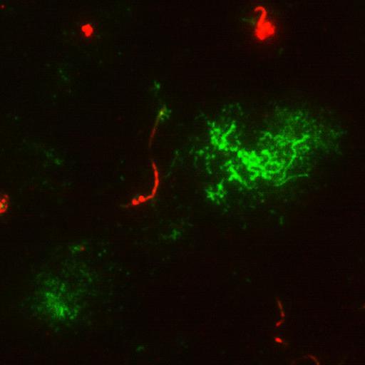
Total internal reflection (TIRF) micrograph of MDCK cells stably transfected with the apical membrane protein P75-GFP (green) and stained for acetylated tubulin of the primary cilium (red) by immunofluorescence.

Technical Details

Cells were grown for 5 days prior to imaging with apical TIRF microscopy. Images were taken with a Leica AM TIRF MC. Penetration depth: 90 nmImage collected using a True MultiColor Laser TIRF Leica AM TIRF MC. See http://www.leica-microsystems.com/science-lab/protein-transport-processes-at-the-apical-membrane-of-polarized-epithelial-cells-applications-for-tirf-microscopy/ for additional details.

Biological Sources
NCBI Organism Classification
Canis lupus familiaris
Cell Type
epithelial cell
Cell Line
MDCK-II
Cellular Component
primary cilium
acetylated tubulin
P-75
Attribution
Names
Christoph Greb
Ralf Jacob
Anja Schu
Citation
Digital Object Identifier (DOI)
doi:10.7295/W9CIL41313
Archival Resource Key (ARK)
ark:/b7295/w9cil41313
Imaging
Image Type
recorded image
Image Mode
total internal reflection microscopy (TIRF)
Parameters Imaged
fluorescence emission
Source of Contrast
distribution of epitope
distribution of a specific protein
Visualization Methods
EGFP
primary antibody plus labeled secondary antibody
Sample Preparation
Methods
living tissue
Relation To Intact Cell
dispersed cells in vitro
Dimensions
Spatial Axis Image Size Pixel Size
X 709px ——
Y 708px ——