Licensing

Pd
Public Domain: This image is in the public domain and thus free of any copyright restrictions. However, as is the norm in scientific publishing and as a matter of courtesy, any user should credit the content provider for any public or private use of this image whenever possible. Learn more
Comments: Powered by Pnyxe

Description

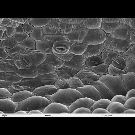
Scanning electron microscope image of a Hibiscus schizopetalus anther. Image shows open and closed stomata (pores on the surface of plant epidermis) and is a high magnification of CIL 41305.

Technical Details

Image collected on a Zeiss DSM 962 SEM. See detailed protocol at http://www.dartmouth.edu/~emlab/manuals/sempreps/index.html

Biological Sources

NCBI Organism Classification
Hibiscus schizopetalus
Cellular Component
stomata

Biological Context

Biological Process
regulation of respiratory gaseous exchange
Plant Growth
anther

Attribution

Names
Louisa Howard
Charles Daghlian
Link
Dartmouth EM Facility

Grouping

This image is part of a group.

Imaging

Image Type
recorded image
Imaging Mode
scanning electron microscopy (SEM)
Parameters Imaged
elastic scattering of electrons
Source of Contrast
differences in deposition of metal shadow
Visualization Methods
shadowing and plating
Processing History
unprocessed raw data

Sample Preparation

Methods
glutaraldehyde fixed tissue
formaldehyde fixed tissue
ethanol dehydration
Relation To Intact Cell
whole mounted tissue

Dimensions

Spatial Axis Image Size Pixel Size
X 1024px ——
Y 749px ——
*CIL – Cell Image Library accession number. Please use this to reference an image.