Alternate header for print version


Licensing
Attribution Non-Commercial Share Alike:This image is licensed under a Creative Commons Attribution, Non-Commercial Share Alike License. View License Deed | View Legal Code
tweet  
*CIL – Cell Image Library accession number. Please use this to reference an image.

CIL:41041*  Cite 
Description

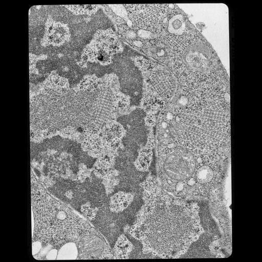
Transmission electron micrograph of a section through a 3-day old chick embryo obtained from an egg that was maintained in a cold room for 24 hrs. Ribosomes have crystallized both in the cytoplasm and nucleus.

Technical Details

See: T. Morimoto et al. 1972. Ribosome crystallization in chicken embryos. I. Isolation, characterization, and in vitro activity of ribosome tetramers. J Cell Biol 52:355-366. B. Byers 1966. Ribosome crystallization induced in chick embryo tissues by hypothermia. J Cell Biol 30:C1-6. Original 3.25 in. x 4 in. lantern slides were scanned at 600dpi

Biological Sources
NCBI Organism Classification
Gallus gallus gallus
Cell Type
early embryonic cell
Cellular Component
nucleus
cytoplasm
ribosome
ribosome crystal
Biological Context
Biological Process
cytoplasm organization
Molecular Function
nucleus organization
Attribution
Names
George E. Palade
Gunter Blobel
T. Morimoto
Link
George E. Palade EM Slide Collection
Citation
Digital Object Identifier (DOI)
doi:10.7295/W9CIL41041
Archival Resource Key (ARK)
ark:/b7295/w9cil41041
Grouping This image is part of a group.
Imaging
Image Type
film
Image Mode
transmission electron microscopy (TEM)
Parameters Imaged
electron density
Source of Contrast
differences in adsorption or binding of stain
Visualization Methods
uranyl salt
Data Qualifiers
processed data
Sample Preparation
Methods
chemically fixed tissue
tissue in plastic embedment
Relation To Intact Cell
microtome-sectioned tissue
Dimensions
Spatial Axis Image Size Pixel Size
X 3584px ——
Y 4528px ——