Alternate header for print version


Licensing
Public Domain: This image is in the public domain and thus free of any copyright restrictions. However, as is the norm in scientific publishing and as a matter of courtesy, any user should credit the content provider for any public or private use of this image whenever possible. Learn more
tweet  
*CIL – Cell Image Library accession number. Please use this to reference an image.

CIL:40968*  Cite 
Description

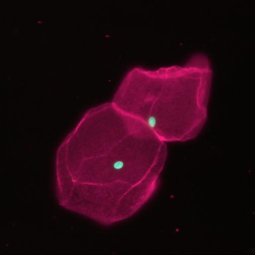
Fluorescent micrograph of epithelial cells from the bladder wall. The cells were isolated immediately after collection of a urine specimen and are not fixed. The image was taken using a standard epi-fluorescent microscope.

Biological Sources
NCBI Organism Classification
Homo sapiens
Cell Type
epithelial cell
Cellular Component
plasma membrane
nucleus
Biological Context
Biological Process
nucleus localization
Human Development Anatomy
bladder
Attribution
Names
Arjan Tibbe
Citation
Digital Object Identifier (DOI)
doi:10.7295/W9CIL40968
Archival Resource Key (ARK)
ark:/b7295/w9cil40968
Imaging
Image Type
recorded image
Image Mode
fluorescence microscopy
Parameters Imaged
fluorescence emission
Source of Contrast
distribution of epitope
Visualization Methods
acridine orange
CellMask Deep Red (Invitrogen)
Processing History
unprocessed raw data
Sample Preparation
Methods
unfixed tissue
Relation To Intact Cell
isolated from human urine sample
Dimensions
Spatial Axis Image Size Pixel Size
X 1024px ——
Y 1022px ——