Alternate header for print version


Licensing
Attribution Non-Commercial; No Derivatives:This image is licensed under a Creative Commons Attribution, Non-Commercial, No Derivatives License. View License Deed | View Legal Code
tweet  
*CIL – Cell Image Library accession number. Please use this to reference an image.

CIL:40967*  Cite 
Description

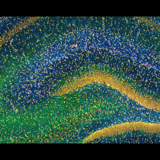
Widefield multiphoton fluorescence image of Rat hippocampus stained to reveal the distribution of glia (cyan), neurofilaments (green) and cell nuclei (yellow). 2nd Prize, 2010 Olympus BioScapes Digital Imaging Competition®.

Technical Details

Biological Sources
NCBI Organism Classification
Rattus
Cell Type
glial cell
neuron
Cellular Component
neurofilament cytoskeleton
nucleus
Biological Context
Biological Process
hippocampus development
Mouse Gross Anatomy
TS25,hippocampus,cerebral cortex
Attribution
Names
Thomas Deerinck
2010 Olympus BioScapes Digital Imaging Competition®
Link
2010 Olympus BioScapes Digital Imaging Compe...
OTHER
National Center for Microscopy and Imaging Research,
University of California, San Diego, CA, USA
Citation
Digital Object Identifier (DOI)
doi:10.7295/W9CIL40967
Archival Resource Key (ARK)
ark:/b7295/w9cil40967
Imaging
Image Type
recorded image
Image Mode
multi-photon microscopy
Parameters Imaged
fluorescence emission
Source of Contrast
distribution of epitope
Visualization Methods
primary antibody plus labeled secondary antibody
Sample Preparation
Relation To Intact Cell
sectioned tissue
Dimensions
Spatial Axis Image Size Pixel Size
X 3600px ——
Y 2857px ——