Alternate header for print version


Licensing
Public Domain: This image is in the public domain and thus free of any copyright restrictions. However, as is the norm in scientific publishing and as a matter of courtesy, any user should credit the content provider for any public or private use of this image whenever possible. Learn more
tweet  
*CIL – Cell Image Library accession number. Please use this to reference an image.

CIL:40903*  Cite 
Description

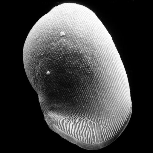
Conchophthirus was deciliated by calcium ion shock followed by shearing through a micropipette. This revealed the location of the deciliated basal bodies and the cell's surface architecture. In this image of the left side ciliature the thigmotactic ciliature is revealed at the suture line. Also note that, when zoomed in, in this preparation the clathrin coated pits, which are normally an invagination into the cytoplasm have everted following the deciliation procedure, to form a small evagination anterior to the stubb of each deciliated basal body. For more information see: Antipa, G. A. and Small, E. B. 1971. A redescription of Conchophthirus curtus Engelmann, 1862 (Protozoa, Ciliatea). J. Protozool. 18:491-503. This micrograph was taken in 1968 by G. Antipa on a Cambridge Mark IIA operating at 20kV. The negative magnification is 765X. The raw film was scanned with an Epson Perfection V750 Pro. This image is available for quantitative analysis.

Biological Sources
NCBI Organism Classification
uncultured scuticociliate
Conchophthirus curtus
Cell Type
cell by organism
eukaryotic cell
Eukaryotic Protist
Ciliated Protist
Cellular Component
cilium
caudal cilia
coated pit
Biological Context
Biological Process
cortical cytoskeleton organization
Attribution
Names
Gregory Antipa (San Francisco State University)
Citation
Digital Object Identifier (DOI)
doi:10.7295/W9CIL40903
Archival Resource Key (ARK)
ark:/b7295/w9cil40903
Grouping This image is part of a group.
Imaging
Image Type
recorded image
Image Mode
illumination by electrons
secondary_electron imaging
Parameters Imaged
illumination by electrons
secondary electron generation
secondary_electron imaging
Source of Contrast
detection of electrons
Visualization Methods
Deciliation
Processing History
recorded image
film
Data Qualifiers
raw, unprocessed data
suitable for spatial measurements
Sample Preparation
Methods
shadowing and plating
chemically fixed tissue
Parducz fixative
lyophilized specimen
Au/Pd metal sputter-coat
Relation To Intact Cell
whole mounted tissue
Dimensions
Spatial Axis Image Size Pixel Size
X 2991px 17nm
Y 4000px 17nm