Alternate header for print version


Licensing
Public Domain: This image is in the public domain and thus free of any copyright restrictions. However, as is the norm in scientific publishing and as a matter of courtesy, any user should credit the content provider for any public or private use of this image whenever possible. Learn more
tweet  
*CIL – Cell Image Library accession number. Please use this to reference an image.

CIL:40693*  Cite 
Description

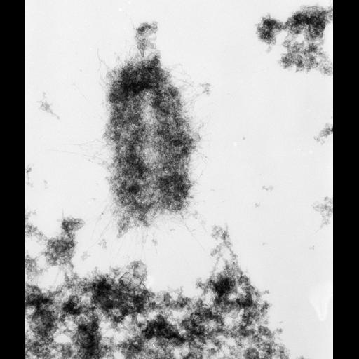
High voltage (1MeV) transmission electron microscopy image of an isolated metaphase chromatid pair from a mouse A9 cell, showing the fibrous structure. The image was taken with a specimen tilt of 45 degrees. Grouped with it is an image with a tilt of 55 degrees, providing a pair that affords an oblique stereo view of the chromosome.

Technical Details

Chromosomes were isolated from metaphase cells using a non-aqueous protocol, exposed to non-ionic detergent for 2 hr, then centrifuged through a sucrose cushion containing formaldehyde onto a carbon-coated grid (spreading technique of O.L. Miller), and critical point dried. See also: H. Ris 1981 Stereoscopic electron microscopy of chromosomes. Meth Cell Biol 22:77-96 H. Ris 1978 Preparation of chromatin and chromosomes for electron microscopy. Meth Cell Biol 18:220-246.

Biological Sources
NCBI Organism Classification
Mus musculus
Cell Type
fibroblast
Cell Line
A9
Cellular Component
nuclear chromosome
Biological Context
Biological Process
chromosome organization
mitosis
mitotic metaphase
Attribution
Names
Hans Ris
Citation
Digital Object Identifier (DOI)
doi:10.7295/W9CIL40693
Archival Resource Key (ARK)
ark:/b7295/w9cil40693
Grouping This image is part of a group.
Imaging
Image Type
film
Image Mode
Parameters Imaged
electron density
Source of Contrast
differences in adsorption or binding of stain
Visualization Methods
uranyl salt
Data Qualifiers
raw, unprocessed data
suitable for spatial measurements
Dimensions
Spatial Axis Image Size Pixel Size
X 4394px 1.58nm
Y 5120px 1.58nm