Alternate header for print version


Licensing
This image is copyright protected. Any public or private use of this image is subject to prevailing copyright laws. Please contact the content provider of this image for permission requests.
tweet  
*CIL – Cell Image Library accession number. Please use this to reference an image.

CIL:40651*  Cite 
Description

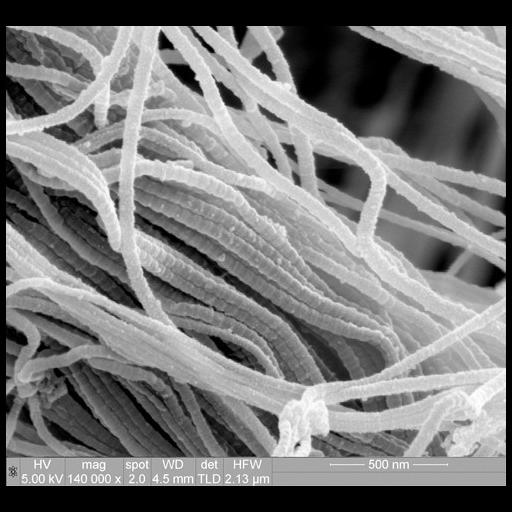
Scanning electron micrograph of collagen fibrils, showing their characteristic 'D banding' morphology. These collagen fibrils are present in the joint capsule tissue that surrounds the knee.

Technical Details

Image collected on a FEI instrument: Quanta Family using Magnification: 140000x, Horizontal Field Width: 2.13 μm, Voltage: 5.00 kV, Detector: TLD, Spot: 2.0 nA, and Working Distance: 4.5 mm.

Biological Sources
Cellular Component
fibrillar collagen
Biological Context
Biological Process
limb joint morphogenesis
Human Development Anatomy
knee
knee joint primordium
Attribution
Names
Paul Gunning
Link
FEI Life Science Images
OTHER
Smith & Nephew
Citation
Digital Object Identifier (DOI)
doi:10.7295/W9CIL40651
Archival Resource Key (ARK)
ark:/b7295/w9cil40651
Imaging
Image Type
recorded image
Image Mode
scanning electron microscopy (SEM)
Parameters Imaged
elastic scattering of electrons
Processing History
unprocessed raw data
Sample Preparation
Relation To Intact Cell
whole mounted tissue
Dimensions
Spatial Axis Image Size Pixel Size
X 500px ——
Y 460px ——