Licensing

Pd
Public Domain: This image is in the public domain and thus free of any copyright restrictions. However, as is the norm in scientific publishing and as a matter of courtesy, any user should credit the content provider for any public or private use of this image whenever possible. Learn more
Comments: Powered by Pnyxe

Description

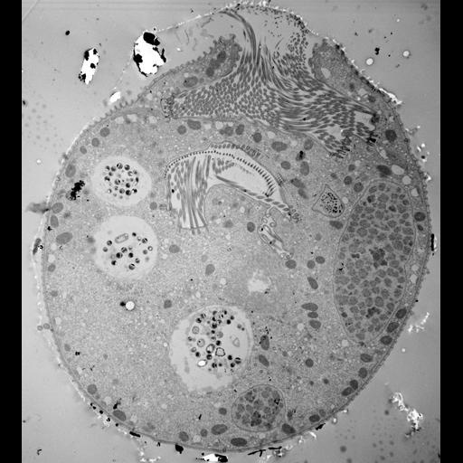
The 11 micrographs of the group are views of a serially-sectioned contracted Vorticella convallaria cell that show the main features of this cell. This figure shows 3 food vacuoles; 2 segments of the macronucleus; micronucleus; perioral cilia; tips of “ribbed wall” along adoral side of the peristome that contain the lamellae; paroral cilia; contractile vacuole; tube or chamber containing bacteria linking the contractile vacuole to the peristome; and myonemes linking the perioral zone to the cell's lateral pellicle. TEM taken on 4/1/71 by R. Allen with Hitachi HU11A operating at 75kV. Neg. 2,150X. The raw negative was scanned with an Epson Perfection V750 Pro and this high resolution image is best used for quantitative analysis. Additional information available at (http://www5.pbrc.hawaii.edu/allen/).

Technical Details

Standard glutaraldehyde fixation followed by osmium tetroxide, dehydrated in alcohol and embedded in an epoxy resin. Microtome sections prepared at approximately 75nm thickness. Additional information available at (http://www5.pbrc.hawaii.edu/allen/).

Biological Sources

NCBI Organism Classification
Vorticella convallaria
Cell Type
cell by organism
eukaryotic cell
Eukaryotic Protist
Ciliated Protist
Cell Line
Cambridge Culture Collection, Cambridge, England, # LB1690/1
Cellular Component
cell
food vacuole
macronucleus
micronucleus
oral apparatus
contractile vacuole

Attribution

Name
Richard Allen (University of Hawaii)
Link
http://www5.pbrc.hawaii.edu/allen/

Grouping

This image is part of a group.

Imaging

Image Type
transmission electron microscopy (TEM)
illumination by electrons
Imaging Mode
detection of electrons
film
Parameters Imaged
electron density
Source of Contrast
stain with broad specificity
Visualization Methods
stain with broad specificity
osmium tetroxide
uranyl salt
lead salt
Processing History
recorded image
film
Print from negative scanned for Photoshop.
Data Qualifiers
processed data

Dimensions

Spatial Axis Image Size Pixel Size
X 3624px 3.7nm
Y 4000px 3.7nm
*CIL – Cell Image Library accession number. Please use this to reference an image.