Alternate header for print version


Licensing
This image is copyright protected. Any public or private use of this image is subject to prevailing copyright laws. Please contact the content provider of this image for permission requests.
tweet  
*CIL – Cell Image Library accession number. Please use this to reference an image.

CIL:40620*  Cite 
Description

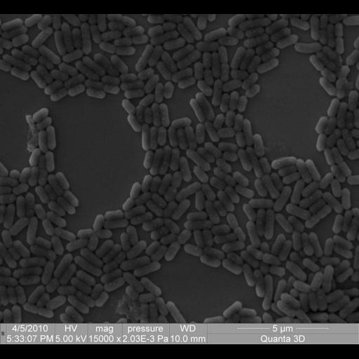
Scanning electron micrograph of Listeria Bacteria. Listeria cause listeriosis, a serious infection in humans induced by eating food contaminated with the bacteria. This image was collected as part of an analysis of qualitative and quantitative changes in a sample containing bacteria,

Technical Details

Image collected using a Quanta 200 3D from the FEI Quanta DualBeam Family. The magnification is 15,000x, the Vacuum was 2,03E-3 Pa, and the voltage was 5 kV, An ETD Spot detector with a 2,5 nA at a working distance of 10 mm was used to collect the data.

Biological Sources
NCBI Organism Classification
Listeria
Cellular Component
cell surface
Biological Context
Biological Process
detection of bacterium
Attribution
Names
Natalia Shevlyagina
Link
FEI Life Sciences Images
Citation
Digital Object Identifier (DOI)
doi:10.7295/W9CIL40620
Archival Resource Key (ARK)
ark:/b7295/w9cil40620
Imaging
Image Type
recorded image
Image Mode
scanning electron microscopy (SEM)
Parameters Imaged
elastic scattering
Source of Contrast
differences in deposition of metal shadow
Visualization Methods
visualization of contiguous regions
Processing History
unprocessed raw data
Sample Preparation
Relation To Intact Cell
dispersed cells in vitro
Dimensions
Spatial Axis Image Size Pixel Size
X 670px ——
Y 617px ——