Alternate header for print version


Licensing
This image is copyright protected. Any public or private use of this image is subject to prevailing copyright laws. Please contact the content provider of this image for permission requests.
tweet  
*CIL – Cell Image Library accession number. Please use this to reference an image.

CIL:40606*  Cite 
Description

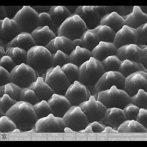
Scanning electron micrograph of lotus leaf trichomes. Trichomes are derived from specialized epidermal cells on a leaf surface and are important for water repellence.

Technical Details

Image collected on a FEI Quanta DualBeam Family Instrument using the ollowing parameters: Magnification: 1500x, Horizontal Field Width: 20um, Vacuum: 10^-6 Torr, Voltage: 30 kV, Detector: ETD, Spot: 3.0 nA, and Working Distance: 10 mm.

Biological Sources
NCBI Organism Classification
Nelumbo nucifera
Cell Type
epidermal cell
Cellular Component
trichome
Biological Context
Biological Process
water repellence
Plant Growth
leaf
Attribution
Names
Craig Queenan
Link
FEI Life Sciences Images
Citation
Digital Object Identifier (DOI)
doi:10.7295/W9CIL40606
Archival Resource Key (ARK)
ark:/b7295/w9cil40606
Imaging
Image Type
recorded image
Image Mode
scanning electron microscopy (SEM)
Parameters Imaged
elastic scattering of electrons
Processing History
unprocessed raw data
Sample Preparation
Relation To Intact Cell
whole mounted tissue
Dimensions
Spatial Axis Image Size Pixel Size
X 670px ——
Y 617px ——