Licensing

Copyright
This image is copyright protected. Any public or private use of this image is subject to prevailing copyright laws. Please contact the content provider of this image for permission requests.
Comments: Powered by Pnyxe

Description

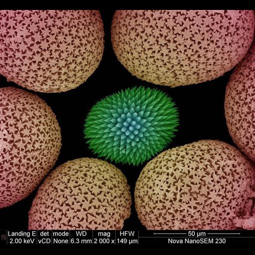
This colorized scanning electron micrograph shows uncoated sponge spicules from a South African sponge. Spicules provide structural support and deter predators. The image was taken with the VcD (backscatter) detector, using beam decelleration

Technical Details

Image collected on FEI instrument: Nova NanoSEM Family using Magnification: 2,000x Horizontal Field Width: 149 μm Voltage: 2 kV Detector: vcd Spot: 2 nA Working Distance: 6.3mm

Biological Sources

NCBI Organism Classification
Porifera
(sponges)
Cellular Component
cell surface
spicule

Biological Context

Biological Process
structural support
deter predator

Attribution

Name
Miranda Waldron
Link
FEI Life Science Images

Imaging

Image Type
recorded image
Imaging Mode
scanning electron microscopy (SEM)
Parameters Imaged
secondary electron generation
Visualization Methods
visualization of contiguous regions
Processing History
unprocessed raw data

Sample Preparation

Relation To Intact Cell
whole mounted tissue

Dimensions

Spatial Axis Image Size Pixel Size
X 670px ——
Y 617px ——
*CIL – Cell Image Library accession number. Please use this to reference an image.