Alternate header for print version


Licensing
This image is copyright protected. Any public or private use of this image is subject to prevailing copyright laws. Please contact the content provider of this image for permission requests.
tweet  
*CIL – Cell Image Library accession number. Please use this to reference an image.

CIL:40580*  Cite 
Description

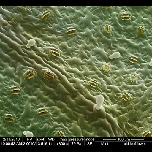
Colorized Scanning electron micrograph of an unfixed mint leaf showing stomata pores.

Technical Details

Image collected on an FEI instrument: Quanta Family using Magnification: 800x, Horizontal Field Width: 320um, Vacuum: 79Pa, Voltage: 2.0 kV, Detector: LFD, Spot: 3.5 nA, and Working Distance: 6.1mm.

Biological Sources
NCBI Organism Classification
Lamiaceae
Cell Type
epidermal cell
Cellular Component
stomata
Biological Context
Biological Process
respiratory gaseous exchange
regulation of water loss via skin
Attribution
Names
David McCarthy
Link
FEI Life Science Images
Citation
Digital Object Identifier (DOI)
doi:10.7295/W9CIL40580
Archival Resource Key (ARK)
ark:/b7295/w9cil40580
Imaging
Image Type
recorded image
Image Mode
scanning electron microscopy (SEM)
Parameters Imaged
elastic scattering of electrons
Processing History
colorized
Sample Preparation
Methods
unfixed tissue
Relation To Intact Cell
whole mounted tissue
Dimensions
Spatial Axis Image Size Pixel Size
X 670px ——
Y 617px ——