Alternate header for print version


Licensing
Attribution Non-Commercial Share Alike:This image is licensed under a Creative Commons Attribution, Non-Commercial Share Alike License. View License Deed | View Legal Code
tweet  
*CIL – Cell Image Library accession number. Please use this to reference an image.

CIL:40560*  Cite 
Description

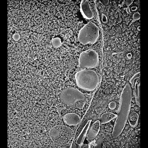
Higher magnification of acidosomes docked at the nascent digestive vacuole membrane. The E-fracture face is relatively IMP free while the P-fracture face has many large IMPs as well as some sizable pits where chunks of this leaflet seem to have stuck to the E-face. Tiny pits on the E-face indicate the removal of individual IMPs from this leaflet. An indentation of the membrane into the lumen of the acidosome shows IMPs on its exposed P-face; which, if any, of these IMPs are V-ATPases is not known. TEM taken on 5/26/92 by R. Allen with Zeiss 10A operating at 80kV. Neg. 19,800X. Published in J. Cell Sci. 106:411-422, 1993. Adapted with permission.

Technical Details

The raw negative was scanned with an Epson Perfection V750 Pro and this high resolution image is best used for quantitative analysis. Additional information available at (http://www5.pbrc.hawaii.edu/allen/).

Biological Sources
NCBI Organism Classification
Paramecium multimicronucleatum
Cell Type
cell by organism
eukaryotic cell
Eukaryotic Protist
Ciliated Protist
Cellular Component
early phagosome
vesicle membrane
oral apparatus
Biological Context
Biological Process
membrane docking
phagosome acidification
digestive system process
Attribution
Names
Richard Allen (University of Hawaii)
Published
J Cell Sci. 1993 Sep;106 ( Pt 1):411-22.
Pubmed
8270639
Citation
Digital Object Identifier (DOI)
doi:10.7295/W9CIL40560
Archival Resource Key (ARK)
ark:/b7295/w9cil40560
Grouping This image is part of a group.
Imaging
Image Type
transmission electron microscopy (TEM)
illumination by electrons
Image Mode
electron density
Parameters Imaged
elastic scattering of electrons
electron density
elevation
Source of Contrast
differences in deposition of metal shadow
Visualization Methods
transmission electron microscopy (TEM)
Processing History
freeze_fracture/freeze_etch
shadowing and plating
recorded image
film
Data Qualifiers
raw, unprocessed data
suitable for spatial measurements
Dimensions
Spatial Axis Image Size Pixel Size
X 4976px 0.75nm
Y 5554px 0.75nm