By 5-8 minutes the membrane of the DV uniformly labels with the mAb to the L1 antigen that is specific for acidosomes and phagoacidosome (DV-II) membrane. An unlabeled lysosome lies next to the DV-II. TEM taken on 11/30/92 by R. Allen with Zeiss 10A operating at 80kV. Neg.19,800X. Published in J. Cell Sci. 108:1263-1274, 1995. Adapted with permission.
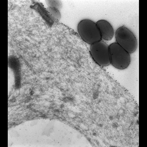
To label membranes inside the cell we used very lightly fixed cells (0.25% glutaraldehyde) that were then rapidly frozen in liquid nitrogen and sectioned later at -100oC. These frozen sections were picked up on drops of methylcellulose and transferred to a Formvar-supported grid. The sections were immunogold labeled (15nm gold) to show the location of the specific antigen inside the cell as well as on the cell surface. Microtome sections prepared at approximately 75nm thickness. The raw negative was scanned with an Epson Perfection V750 Pro and this high resolution image is best used for quantitative analysis. Additional information available at (http://www5.pbrc.hawaii.edu/allen/).
| Spatial Axis | Image Size | Pixel Size |
|---|---|---|
| X | 5047px | 0.75nm |
| Y | 5649px | 0.75nm |