Alternate header for print version


Licensing
Attribution Non-Commercial Share Alike:This image is licensed under a Creative Commons Attribution, Non-Commercial Share Alike License. View License Deed | View Legal Code
tweet  
*CIL – Cell Image Library accession number. Please use this to reference an image.

CIL:40444*  Cite 
Description

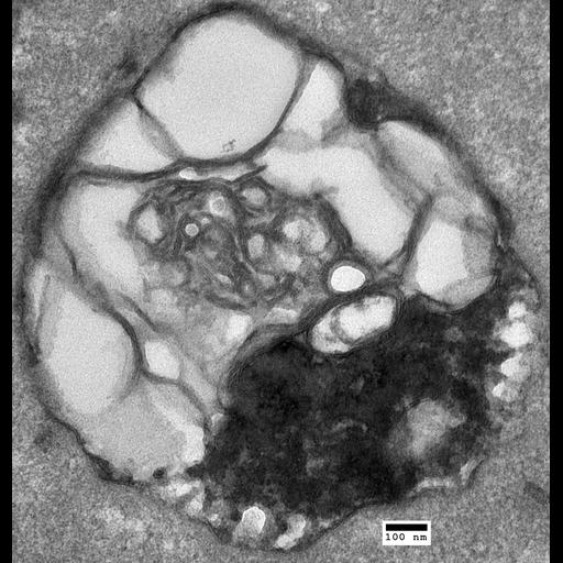
Transmission electron micrograph of an autophagolysosome from a sertraline-treated prototrophic wildtype cell. (20,000X). This image is part of a group of yeast vacuole images (CIL:40437-40448).

Technical Details

This group of images contains representative whole-cell images and high-magnification zooms of organelle structures of interest. All cells were grown in liquid rich media (YPD pH6.5) at 30C with aerated shaking, and cultures were sampled in mid-logarithmic phase. Treated cells were exposed to 60µM antidepressant sertraline (Zoloft®) for 45 minutes.

Biological Sources
NCBI Organism Classification
Saccharomyces cerevisiae
Cellular Component
autophagolysosome
Biological Context
Biological Process
organelle organization
sertraline treatment
Attribution
Names
Ethan Perlstein
Daniel Korostyshevsky
OTHER
Princeton EM Core Facility
Citation
Digital Object Identifier (DOI)
doi:10.7295/W9CIL40444
Archival Resource Key (ARK)
ark:/b7295/w9cil40444
Grouping This image is part of a group.
Imaging
Image Type
recorded image
Image Mode
transmission electron microscopy (TEM)
Parameters Imaged
electron density
Source of Contrast
differences in adsorption or binding of stain
Visualization Methods
osmium tetroxide
Processing History
unprocessed raw data
Sample Preparation
Methods
glutaraldehyde fixed tissue
tissue in plastic embedment
Relation To Intact Cell
sectioned tissue
Dimensions
Spatial Axis Image Size Pixel Size
X 2440px ——
Y 2556px ——