Alternate header for print version


Licensing
Attribution Non-Commercial Share Alike:This image is licensed under a Creative Commons Attribution, Non-Commercial Share Alike License. View License Deed | View Legal Code
tweet  
*CIL – Cell Image Library accession number. Please use this to reference an image.

CIL:40316*  Cite 
Description

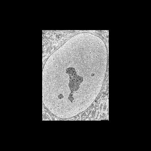
Transmission electron micrograph of a thin section of the nucleus of a normal HeLa cell - from a study of the role of the nuclear porin ALADIN. Other images in this group show details of nuclear pore structure in cross section and tangential section.

Technical Details

Normal HeLa cells expressing were fixed with glutaraldehyde, embedded in epoxy resin, thin sectioned, and imaged using TEM. See: J Cronshaw and M Matunis 2003 The nuclear pore complex ALADIN is mislocalized in triple A syndrome Proc Natl Acad Sci USA 100 5823-5827

Biological Sources
NCBI Organism Classification
Homo sapiens
Cell Type
epithelial cell
cervical carcinoma
Cell Line
HeLa
Cellular Component
nucleus
nuclear envelope
nuclear chromatin
nucleolus
Biological Context
Biological Process
nucleus organization
nuclear transport
Attribution
Names
Janet Cronshaw
Michael Matunis
Published
J Cronshaw and M Matunis 2003 The nuclear pore complex ALADIN is mislocalized in triple A syndrome Proc Natl Acad Sci USA 100 5823-5827
Pubmed
12730363
Link
Proceedings National Academy of Sciences USA
Citation
Digital Object Identifier (DOI)
doi:10.7295/W9CIL40316
Archival Resource Key (ARK)
ark:/b7295/w9cil40316
Grouping This image is part of a group.
Imaging
Image Type
recorded image
Image Mode
transmission electron microscopy (TEM)
Parameters Imaged
electron density
Source of Contrast
differences in adsorption or binding of stain
Visualization Methods
osmium tetroxide
uranyl salt
lead salt
Data Qualifiers
processed data
Sample Preparation
Methods
glutaraldehyde fixed tissue
tissue in plastic embedment
Relation To Intact Cell
microtome-sectioned tissue
Dimensions
Spatial Axis Image Size Pixel Size
X 226px ——
Y 307px ——