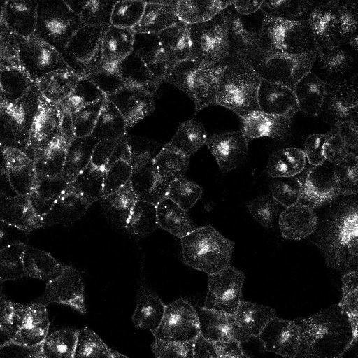
Maximum intensity projection of a confocal z series of MDCK cells expressing Cx43-GFP-4C imaged with confocal microscopy. This image has been downsampled from the raw data image, and time lapse series, both of which can be accessed using the link provided to the Cell Centered Database. For more information, see: Boassa et al. (2010) Trafficking and Recycling of the Connexin43 Gap Junction Protein during Mitosis. Traffic. PMID: 20716111.
Madin-Darby canine kidney (MDCK) cells were maintained at 37° C, and 10% CO2 in Dulbecco's modified Eagle's medium containing 10% fetal bovine serum (FBS) (Gibco-BRL, Invitrogen). Transductions were carried out using a retroviral system according to the protocols from the Nolan laboratory (www.stanford.edu/group/nolan). Experiments were conducted on endogenous expressing or stably Cx43-expressing cell lines generated by transduction followed by selection with the antibiotic hygromycin (Gibco-BRL, Invitrogen). In order to image several cells progressing through mitosis, cells were synchronized using a combination of serum starvation and application of aphidicolin (APD), an inhibitor of DNA synthesis, to create an enriched population of cells undergoing mitosis. Confluent cells were trypsinized and grown for about 20 h in the presence of minimal serum and APD, resulting in a G1/S block. The drug was then washed out, normal serum was restored and the cells were allowed to progress through S phase to mitosis. Samples were mounted in Opti-MEM and time-lapse imaging of the intrinsic GFP was performed after 7 h, when cells began entering mitosis, using an Olympus Fluoview 1000 with a 60X NA 1.42 objective.
| Spatial Axis | Image Size | Pixel Size |
|---|---|---|
| X | 512px | —— |
| X | 512px | —— |