Alternate header for print version


Licensing
Attribution Only: This image is licensed under a Creative Commons Attribution License. View License Deed | View Legal Code
tweet  
*CIL – Cell Image Library accession number. Please use this to reference an image.

CIL:40229*  Cite 
Description

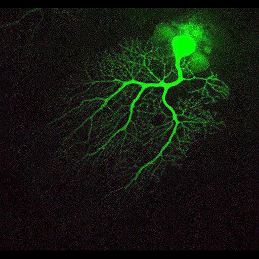
A Purkinje neuron from the cerebellum of a 2 month old male C57BL/6 mouse, injected with Lucifer Yellow. The elaborate, highly branched dendritic arbor is revealed in this maximum projection image which was generated by compiling a z-series of optical sections. This image has been downsampled from the raw data image which can be accessed using the link provided to the Cell Centered Database.

Technical Details

Tissue was perfused with paraformaldehyde, sectioned (100 µm) on a vibratome and mounted in gelvatol, and imaged on a Biorad Radiance 2000 Confocal using a 60x 1.4NA objective.

Biological Sources
NCBI Organism Classification
Mus musculus
Cell Type
Purkinje cell
Cellular Component
dendrite
neuronal cell body
Biological Context
Biological Process
cell projection organization
Attribution
Names
Maryann Martone
Andrea Price
Andrea Thor
Hiro Hakozaki
Masako Terada
Link
CCDB 7
Citation
Digital Object Identifier (DOI)
doi:10.7295/W9CIL40229
Archival Resource Key (ARK)
ark:/b7295/w9cil40229
Grouping This image is part of a group.
Imaging
Image Type
recorded image
Image Mode
confocal microscopy
Parameters Imaged
fluorescence emission
Source of Contrast
intracellular filling
Visualization Methods
lucifer yellow
Data Qualifiers
processed data
Sample Preparation
Relation To Intact Cell
vibratome-sectioned tissue
Dimensions
Spatial Axis Image Size Pixel Size
X 512px ——
Y 533px ——