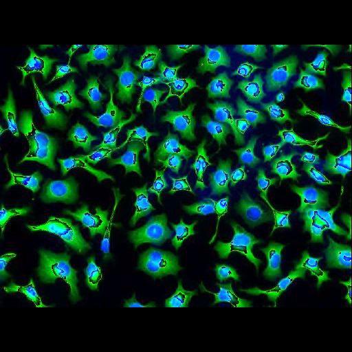
Wide field fluorescent image of cultured neuroblastoma cells labeled with phalloidn FITC (green) and DAPI (blue) nuclear stain. This image has been downsampled from the raw data image which can be accessed using the link provided to the Cell Centered Database. For further information, see Weimiao Yu, Hwee Kuan Lee, Srivats Hariharan, Wen Yu Bu and Sohail Ahmed. Evolving Generalized Voronoi Diagrams of Active Contours for Accurate Cellular Image Segmentation. Cytometry Part A 77A pg. 379~386, 2010.
NIE115 neuroblastoma cells were grown on 18 X 18 mm lamin coated glass coverslips maintained at 37 deg C in 5% CO2. Culture medium was Dulbecoo's Modified Eagle's medium supllemented with 4500 mg/ml glucose and 1% penicillin-streptomycin as an antibiotic. Cells were serum starved for 48 hrs and then fixed with 4% paraformaldehyde, then counterstained with DAPI and FITC-phalloidin. Images were acquired with a Zeiss epifluorescence microscope and a 20X objective.
| Spatial Axis | Image Size | Pixel Size |
|---|---|---|
| X | 512px | —— |
| Y | 382px | —— |