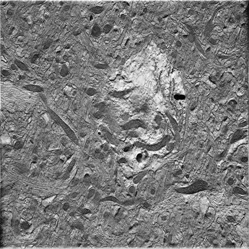
Single computed slice through a tomographic reconstruction of the cell body of a protoplasmic astrocyte in a 0.5 um thick section from the hippocampus of a 1 month old male mouse, imaged with intermediate voltage electron microscopy. This image has been downsampled from the raw data image which can be accessed using the link provided to the Cell Centered Database. The reconstruction is the 21st in a series of 26 serial reconstructions through the cell soma. All 26 images can be found in this image group within the Cell Library, and the complete reconstruction can be viewed in the Cell centered database, accession # 7503.
Detailed methods can be found at the CCDB link provided. Briefly, one month old male mouse (C57BL/6NHsd) was perfused with Ringer's solution for 2 minutes followed by 2% glutaraldehyde and 2% paraformaldehyde in 0.15M sodium cacodylate buffer, pH 7.4. Further dissection of tissue was followed with postfixation using 1% OsO4, additional staining using 2% uranyl acetate, processed for embedding in durcupan ACM resin, and sectioned at a thickness of 5µm thick. Serial tomograms were produced from single tilt images using a JEOL4000, with 8k x 8k CCD camera, 5000X, 400.0 keV).
| Spatial Axis | Image Size | Pixel Size |
|---|---|---|
| X | 512px | —— |
| Y | 512px | —— |