Alternate header for print version


Licensing
Attribution Only: This image is licensed under a Creative Commons Attribution License. View License Deed | View Legal Code
tweet  
*CIL – Cell Image Library accession number. Please use this to reference an image.

CIL:40022*  Cite 
Description

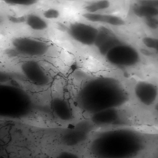
Z projection through a through-focus series of a medium spiny neuron of the neostriatum from a dopamine transporter knock out mouse, injected with Lucifer Yellow and then photoconverted. This image has been downsampled from the raw data image which can be accessed using the link provided to the Cell Centered Database, where the reconstruction of the entire cell can also be found.

Technical Details

Tissue was prepared from a 6 month adult male mouse strain, C57BL/129SvJ. Tissue sections, 100µm, were generated using a vibratome. The entire through series of images were gathered using a Bio-Rad Radiance 2000 with a 40X 0.75 NA.

Biological Sources
NCBI Organism Classification
Mus musculus
Cell Type
CNS neuron (sensu Vertebrata)
Cellular Component
dendrite
axon
Attribution
Names
Diana Price
Link
CCDB 3693
Citation
Digital Object Identifier (DOI)
doi:10.7295/W9CIL40022
Archival Resource Key (ARK)
ark:/b7295/w9cil40022
Imaging
Image Type
recorded image
Image Mode
confocal microscopy
Visualization Methods
intracellular filling
lucifer yellow
Data Qualifiers
processed data
Sample Preparation
Methods
formaldehyde fixed tissue
tissue in epoxy resin embedment
Relation To Intact Cell
microtome-sectioned tissue
Dimensions
Spatial Axis Image Size Pixel Size
X 512px ——
X 512px ——