Alternate header for print version


Licensing
Attribution Only: This image is licensed under a Creative Commons Attribution License. View License Deed | View Legal Code
tweet  
*CIL – Cell Image Library accession number. Please use this to reference an image.

CIL:39960*  Cite 
Description

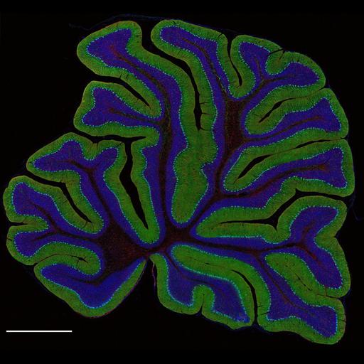
High resolution montage of a sagittal section through the cerebellar cortex of an adult male rat (80g), triple-labeled to reveal nuclei (Hoescht stain, blue), Purkinje neurons (IP3 receptor, green) and astroglia (glial fibrillary acidic protein, GFAP, red). This image is a maximum projection of the Z-stack through the tissue, and has been downsampled from the raw data image which can be accessed using the link provided to the Cell Centered Database.

Technical Details

80µm thick sections were mounted in gelvitol using coverslips with a 1.0 µm thickness. Images were acquired with a BioRad RTS 2000MP Multiphoton microscope using a 40X 1.3NA oil immersion objective. This mosaic image was reconstructed with 10% overlap of individual images; image normalization was performed using ImageJ.

Biological Sources
NCBI Organism Classification
Rattus norvegicus
Cell Type
Purkinje cell
astrocyte
cerebellar granule cell
Cellular Component
cell projection cytoplasm
cell body
nucleus
neuronal cell body
Biological Context
Biological Process
cerebellum structural organization
Attribution
Names
Tom Deerinck
Link
CCDB 23
Citation
Digital Object Identifier (DOI)
doi:10.7295/W9CIL39960
Archival Resource Key (ARK)
ark:/b7295/w9cil39960
Imaging
Image Type
recorded image
Image Mode
multi-photon microscopy
Parameters Imaged
fluorescence emission
Source of Contrast
distribution of epitope
differences in adsorption or binding of stain
Visualization Methods
primary antibody plus labeled secondary antibody
Hoechst 33342
Data Qualifiers
processed data
suitable for spatial measurements
Sample Preparation
Methods
formaldehyde fixed tissue
Relation To Intact Cell
vibratome-sectioned tissue
Dimensions
Spatial Axis Image Size Pixel Size
X 512px 0.36µm
Y 478px 0.36µm