Licensing

Attribution_cc_by
Attribution Only: This image is licensed under a Creative Commons Attribution License. View License Deed | View Legal Code
Comments: Powered by Pnyxe

Description

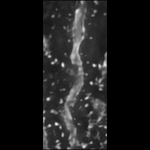
A portion of dendrite and associated spines from a Purkinje neuron of the rat cerebellum. This image is a maximum intensity projection through a computationally deblurred transmitted light series of optical sections. The through focus series was taken from a 2 um section prepared for electron microscopy.

Technical Details

Specimen was imaged using a BioRad MRC 1024 Confocal, using a 63X NA 1.4 objective and 488 laser line.

Biological Sources

NCBI Organism Classification
Rattus norvegicus
(Brown Rat)
Cell Type
CNS neuron (sensu Vertebrata)
Purkinje cell
Cellular Component
dendritic spine
dendrite

Attribution

Names
Maryann Martone
Naoko Yamada
Gordun Arbuthnott
Cali Ingham
Stephen Young
Link
CCDB 15

Imaging

Image Type
recorded image
Imaging Mode
confocal microscopy
Parameters Imaged
fluorescence emission
Data Qualifiers
processed data

Sample Preparation

Relation To Intact Cell
microtome-sectioned tissue

Dimensions

Spatial Axis Image Size Pixel Size
X 564px ——
Y 564px ——
*CIL – Cell Image Library accession number. Please use this to reference an image.