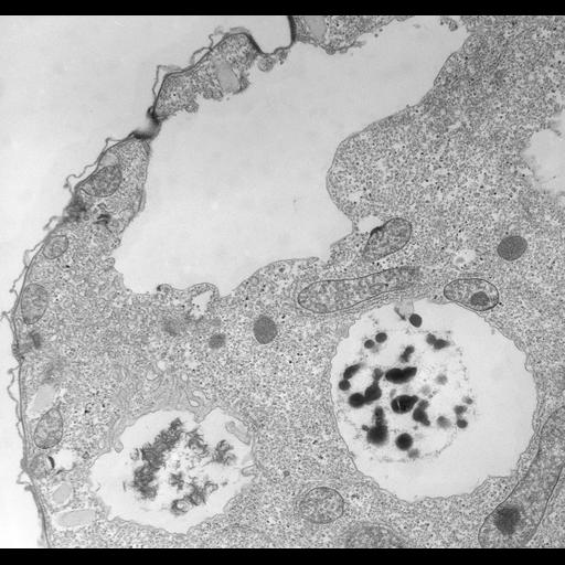
A partially filled contractile vacuole is connected to two regions of spongiome tubules. The extra CV membrane is acquired from these spongiomes. Presumably some of these spongiome tubules are decorated with pegs which in Paramecium are known to be V-type proton pumps. A spent vacuole lies nearby and has a filamentous area in which membrane tubulation and vesiculation is taking place. A similar filamentous array does not appear to be present in the spongiome region of the CV or along the CV membrane itself. TEM taken on 4/21/78 by R. Allen with Hitachi HU11A operating at 60kV. Neg. 9,200X. Bar = 0.5µm. The negative was printed to paper and the image was scanned to Photoshop. This digitized image is available for qualitative analysis. There is a high resolution version of this image in the library (CIL:39799) which is available for quantitative analysis. Additional information available at (http://www5.pbrc.hawaii.edu/allen/).
Standard glutaraldehyde fixation followed by osmium tetroxide, dehydrated in alcohol and embedded in an epoxy resin. Microtome sections prepared at approximately 75nm thickness. Additional information available at (http://www5.pbrc.hawaii.edu/allen/).
| Spatial Axis | Image Size | Pixel Size |
|---|---|---|
| X | 4000px | 1.1nm |
| Y | 3781px | 1.1nm |