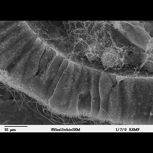
Scanning electron microscope image of Strongylocentrotus drobachiensus [sea urchin] at the gastrula stage. Embryo was split open to reveal a nice cross-section through the outer epithelial layer and the blastocoel cavity. The fertilization envelope dissolves during swimming blastula stage; therefore, cilia and microvilli can be seen at the apical end of the epithelial layer. A few migrating primary mesenchyme cells can be seen within the blastocoel matrix. This is a lower magnification of CIL:39790. This image is part of a group of SEM images of sea urchin embryos at various stages of development. The group is available as CIL:39782-39791.
Images taken on a JEOL 35C SEM. Processing protocol: 1. 2% GTA in 75% MBL SW or FilteredSW FSW - fix for 1.5hrs. RT. 2. Rinse with 0.1M NaCacodylate buffer pH 7.4 3. 0.5% OSO4 in .075M NaCacodylate buffer pH 7.4 - 30' in the cold 4. Rinse in 0.1M NaCacodylate buffer ph 7.4 5. Dehydrate thru ETOH: 30%, 50%, 70%, 95% for 5 min each 6. 100% - 3 changes - 15' each 7. Critical Point Dry 8. Stick embryos down onto silver paint. Gently crack open with glass needle (pipette drawn out to a fine point). 9. Sputter coat with AuPd.
| Spatial Axis | Image Size | Pixel Size |
|---|---|---|
| X | 512px | —— |
| Y | 374px | —— |