Alternate header for print version


Licensing
This image is copyright protected. Any public or private use of this image is subject to prevailing copyright laws. Please contact the content provider of this image for permission requests.
tweet  
*CIL – Cell Image Library accession number. Please use this to reference an image.

CIL:39752*  Cite 
Description

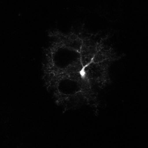
Astrocyte cell from live slices taken from the cortex layer 2/3 of an 8 month old GFAP mouse. In this mouse the enhanced green fluorescent protein (EGFP) under the control of the human glial fibrillary acidic protein (GFAP) promoter (line TgN(GFAP-EGFP)GFEC-FKi). In the image, you can clearly see cell the body, astrocyte cloud and astrocyte projection on vascular tube crossing astrocyte cloud (left and up from cell body). This image an individual z slice. Another z slice is available as CIL 39753, and the projection of 30 slices is available as CIL 30751.

Technical Details

For details on specimen preparation, please see: Palygin O, Lalo U, Verkhratsky A, Pankratov Y. Cell Calcium. 2010 Oct;48(4):225-31 and Lalo U, Palygin O, North RA, Verkhratsky A, Pankratov Y. Aging Cell. 2011 Jun;10(3):392-402. doi: 10.1111/j.1474-9726.2011.00682.x. Epub 2011 Mar 22.

Biological Sources
NCBI Organism Classification
mus musculus
Cell Type
astrocyte
Cellular Component
cell projection
Biological Context
Biological Process
cerebellar cortex structural organization
Attribution
Names
Oleg Palygin
OTHER
Department of Physiology The Medical College of Wisconsin
Citation
Digital Object Identifier (DOI)
doi:10.7295/W9CIL39752
Archival Resource Key (ARK)
ark:/b7295/w9cil39752
Grouping This image is part of a group.
Imaging
Image Type
recorded image
Image Mode
two-photon laser scanning micrograph
Parameters Imaged
fluorescence emission
Source of Contrast
distribution of a specific protein
Visualization Methods
EGFP
Sample Preparation
Methods
brian slices
Relation To Intact Cell
living tissue
Dimensions
Spatial Axis Image Size Pixel Size
X 700px ——
Y 700px ——