Alternate header for print version


Licensing
Public Domain: This image is in the public domain and thus free of any copyright restrictions. However, as is the norm in scientific publishing and as a matter of courtesy, any user should credit the content provider for any public or private use of this image whenever possible. Learn more
tweet  
*CIL – Cell Image Library accession number. Please use this to reference an image.

CIL:39722*  Cite 
Description

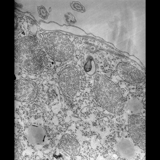
The acidosome-like vesicle has at least 2 enclosing membranes or one membrane indented back on itself and an internal electron-opaque tongue-like droplet that is continuous with the enclosing membranes at one end of the vesicle and may arise by invagination of these membranes into the vesicle’s interior. Such vesicles do not resemble lysosomes of any other organism, including Paramecium. Lysosome membranes should be thicker and be coated by a heavy glycocalyx and exhibit latency. There is no glycocalyx seen here. TEM taken on 8/15/67 by R. Allen with Philips 200 operating at 60kV. Neg. 19,200X. The raw negative was scanned with an Epson Perfection V750 Pro and this high resolution image is best used for quantitative analysis. Additional information available at (http://www5.pbrc.hawaii.edu/allen/).

Technical Details

Standard glutaraldehyde fixation followed by osmium tetroxide, dehydrated in alcohol and embedded in an epoxy resin. Microtome sections prepared at approximately 75nm thickness. Additional information available at (http://www5.pbrc.hawaii.edu/allen/).

Biological Sources
NCBI Organism Classification
Tetrahymena pyriformis
Cell Type
cell by organism
eukaryotic cell
Eukaryotic Protist
Ciliated Protist
Cellular Component
acidosome
organelle membrane
Biological Context
Biological Process
digestive system process
Attribution
Names
Richard Allen (University of Hawaii)
Citation
Digital Object Identifier (DOI)
doi:10.7295/W9CIL39722
Archival Resource Key (ARK)
ark:/b7295/w9cil39722
Grouping This image is part of a group.
Imaging
Image Type
transmission electron microscopy (TEM)
illumination by electrons
Image Mode
detection of electrons
film
Parameters Imaged
electron density
Source of Contrast
stain with broad specificity
Visualization Methods
stain with broad specificity
osmium tetroxide
uranyl salt
lead salt
Processing History
recorded image
film
Data Qualifiers
raw, unprocessed data
suitable for spatial measurements
Dimensions
Spatial Axis Image Size Pixel Size
X 5016px 0.78nm
Y 6118px 0.78nm