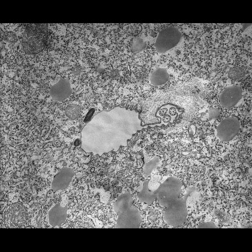
The bottom of the buccal cavity is surrounded by specialized cytoplasm and contains a lip that constricts the opening leading through the cytostome into the receiving vacuole or nascent vacuole. The oral ribs extend along one side of the cytostome and merge into the cytopharynx. Oral ribs have alveoli between the ribs but alveoli stop at the beginning of the cytopharynx. Lamellae, of 2 microtubules each, border the cytopharynx and direct discoidal vesicles to the site of fusion with the membrane of the growing vacuole. Two electron opaque vesicles, called lysosomes by most authors (eg. Nilsson, Biochem. and Phys. of Protozoa, 2nd ed., Vol 2:339-379, 1979, Acad. Press.), which are probably acidosomes, bind to the nascent vacuole membrane. TEM taken on 8/23/67 by R. Allen with Philips 200 operating at 60kV. Neg. 12,400X. The raw negative was scanned with an Epson Perfection V750 Pro and this high resolution image is best used for quantitative analysis. Additional information available at (http://www5.pbrc.hawaii.edu/allen/).
Standard glutaraldehyde fixation followed by osmium tetroxide, dehydrated in alcohol and embedded in an epoxy resin. Microtome sections prepared at approximately 75nm thickness. Additional information available at (http://www5.pbrc.hawaii.edu/allen/).
| Spatial Axis | Image Size | Pixel Size |
|---|---|---|
| X | 6174px | 1.2nm |
| Y | 5038px | 1.2nm |