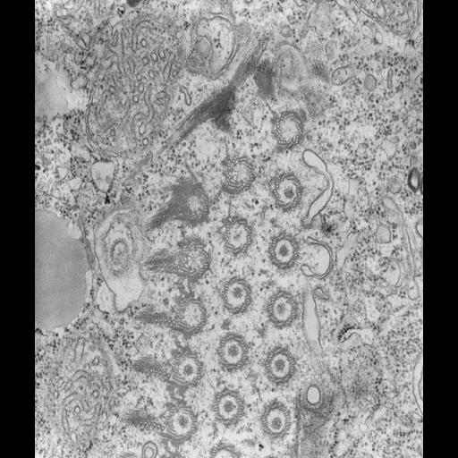
The accessory microtubules at the proximal ends of the polykinetids, both in cross section and tangential section, are highlighted at higher magnification. This section shows one parasomal sac as well as several early endosomes under and to the side of the membranelle. TEM taken on 8/25/67 by R. Allen with Philips 200 operating at 60kV. Neg. 28,000X. The raw negative was scanned with an Epson Perfection V750 Pro and this high resolution image is best used for quantitative analysis. Additional information available at (http://www5.pbrc.hawaii.edu/allen/).
Standard glutaraldehyde fixation followed by osmium tetroxide, dehydrated in alcohol and embedded in an epoxy resin. Microtome sections prepared at approximately 75nm thickness. Additional information available at (http://www5.pbrc.hawaii.edu/allen/).
| Spatial Axis | Image Size | Pixel Size |
|---|---|---|
| X | 3864px | 0.7nm |
| Y | 4561px | 0.7nm |