Alternate header for print version


Licensing
Attribution Non-Commercial; No Derivatives:This image is licensed under a Creative Commons Attribution, Non-Commercial, No Derivatives License. View License Deed | View Legal Code
tweet  
*CIL – Cell Image Library accession number. Please use this to reference an image.

CIL:39473*  Cite 
Description

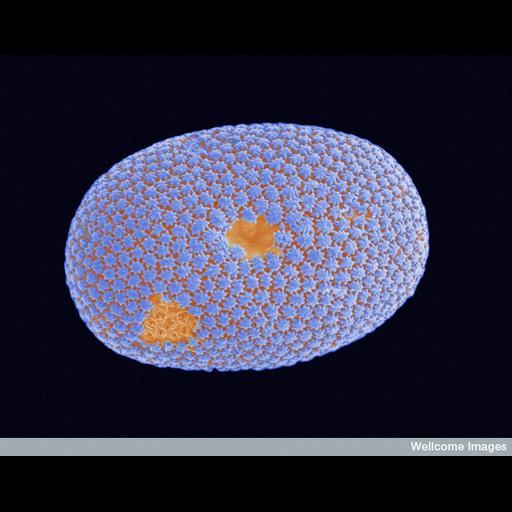
Colorized scanning electron micrograph of a diatom frustule. Diatoms are unicellular organisms and a major group of algae. Diatoms are encased within a hard cell wall made from silica (glass) known as a frustule. Diatom communities are often used to measure environmental conditions, for example water quality.

Technical Details

B0008350 Diatom frustule. Wellcome Images available under the following creative commons usage http://creativecommons.org/licenses/by-nc-nd/2.0/uk/

Biological Sources
NCBI Organism Classification
Bacillariophyta
Cellular Component
cell wall
frustule
Biological Context
Biological Process
unicellular algae organization
Attribution
Names
Anne Weston
Link
Wellcome Images
OTHER
London Research Instiute, CRUK
Citation
Digital Object Identifier (DOI)
doi:10.7295/W9CIL39473
Archival Resource Key (ARK)
ark:/b7295/w9cil39473
Imaging
Image Type
recorded image
Image Mode
scanning electron microscopy (SEM)
Parameters Imaged
elastic scattering of electrons
Source of Contrast
differences in deposition of metal shadow
Visualization Methods
visualization of contiguous regions
Processing History
colorized
Sample Preparation
Relation To Intact Cell
whole mounted tissue
Dimensions
Spatial Axis Image Size Pixel Size
X 734px ——
Y 576px ——