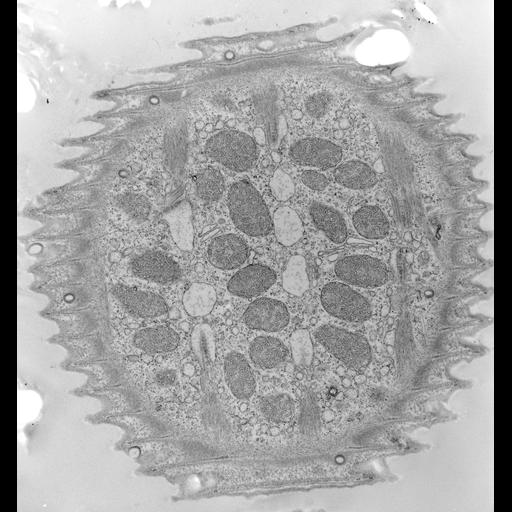
Like other peritrichs the pellicle of Vorticella convallaria is sculpted into ridges and grooves that circle the cell perpendicular to the aboral/adoral axis of the cell body. Pellicular pores penetrate the pellicle as cylindrical indentations of the plasma membrane that end as clathrin-coated pits. A system of alveoli underlies the plasma membrane and rod-like densities occupy the cytosolic tips of the pellicular ridges. A fibrous epiplasm covers the inside of the alveoli. Distinct myonemal bands lie in the aboral to adoral direction to the inside of the epiplasm. These bands are associated with the endoplasmic reticulum by unique specializations of the ER. In this EM preparation the ER has artifactually segmented into vesicles caused by the fixation process. Mitochondria occupy the space between the bands. TEM taken on 4/15/71 by R. Allen with Hitachi HU11A operating at 75kV. Neg. 9,250X. The raw negative was scanned with an Epson Perfection V750 Pro and this high resolution image is best used for quantitative analysis. Additional information available at (http://www5.pbrc.hawaii.edu/allen/).
Standard glutaraldehyde fixation followed by osmium tetroxide, dehydrated in alcohol and embedded in an epoxy resin. Microtome sections prepared at approximately 75nm thickness. Additional information available at (http://www5.pbrc.hawaii.edu/allen/).
| Spatial Axis | Image Size | Pixel Size |
|---|---|---|
| X | 3726px | 1.1nm |
| Y | 4000px | 1.1nm |