Alternate header for print version


Licensing
Attribution Non-Commercial Share Alike:This image is licensed under a Creative Commons Attribution, Non-Commercial Share Alike License. View License Deed | View Legal Code
tweet  
*CIL – Cell Image Library accession number. Please use this to reference an image.

CIL:39461*  Cite 
Description

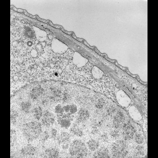
V. convallaria preserved in collidine buffered fixative (not cacodylate) has a less dense cytoplasm in which oranelles are easier to recognize. The rough ER has vesiculated but this vesiculation enhances the identification of loci where ER remains bound to the myonemes. These loci on the ER are called “linkage complexes." These complexes are unique and have not been reported outside the peritrichs. Macronucleus with nucleolus and cytopharynx at its distal end. TEM taken on 3/30/71 by R. Allen with Hitachi HU11A operating at 75kV. Neg. 13,750X. Published and adapted with permission from J. Cell Biol. 56:559-579, 1973. The raw negative was scanned with an Epson Perfection V750 Pro and this high resolution image is best used for quantitative analysis. Additional information available at (http://www5.pbrc.hawaii.edu/allen/).

Technical Details

Standard glutaraldehyde fixation followed by osmium tetroxide, dehydrated in alcohol and embedded in an epoxy resin. Microtome sections prepared at approximately 75nm thickness. Additional information available at (http://www5.pbrc.hawaii.edu/allen/).

Biological Sources
NCBI Organism Classification
Vorticella convallaria
Cell Type
cell by organism
eukaryotic cell
Eukaryotic Protist
Ciliated Protist
Cell Line
Cambridge Culture Collection, Cambridge, England, # LB1690/1
Cellular Component
cell cortex
macronucleus
endoplasmic reticulum
cytopharynx
Attribution
Names
Richard Allen (University of Hawaii)
Published
J Cell Biol. 1973 Feb;56(2):559-79.
Published
4630196
Citation
Digital Object Identifier (DOI)
doi:10.7295/W9CIL39461
Archival Resource Key (ARK)
ark:/b7295/w9cil39461
Grouping This image is part of a group.
Imaging
Image Type
transmission electron microscopy (TEM)
illumination by electrons
Image Mode
detection of electrons
film
Parameters Imaged
electron density
Source of Contrast
stain with broad specificity
Visualization Methods
stain with broad specificity
osmium tetroxide
uranyl salt
lead salt
Processing History
recorded image
film
Data Qualifiers
raw, unprocessed data
suitable for spatial measurements
Dimensions
Spatial Axis Image Size Pixel Size
X 4614px 1.1nm
Y 5308px 1.1nm