Licensing
Description
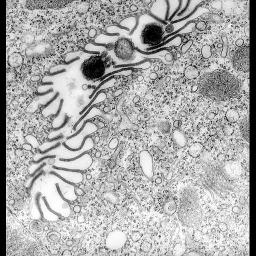
Cytopharynx with numerous folded membrane ribs subtended by five or so microtubules. These microtubules have arms that bind to other microtubules and to the membrane of the cytopharynx and to surrounding vesicles of various sizes. The vesicles are uncoated and free of ribosomes. The vesicles are probably equivalent to the discoidal vesicles and/or to the 100nm vesicles; the latter arise from the endosomal pathway of Paramecium. Evidence for vesicle fusion to the cytopharynx membrane, between the ribs, can be seen. Bacteria and tips of cilia are present in the lumen of the cytopharynx. TEM taken on 4/6/72 by R. Allen with Hitachi HU11A operating at 75kV. Mag. 19,500X. The raw negative was scanned with an Epson Perfection V750 Pro and this high resolution image is best used for quantitative analysis. Additional information available at (http://www5.pbrc.hawaii.edu/allen/).
Technical Details
Standard glutaraldehyde fixation followed by osmium tetroxide, dehydrated in alcohol and embedded in an epoxy resin. Microtome sections prepared at approximately 75nm thickness. Additional information available at (http://www5.pbrc.hawaii.edu/allen/).
Biological Sources
- NCBI Organism Classification
- Vorticella convallaria
- Cell Type
- cell by organism
- eukaryotic cell
- Eukaryotic Protist
- Ciliated Protist
- Cell Line
- Cambridge Culture Collection, Cambridge, England, # LB1690/1
- Cellular Component
- oral apparatus
- microtubule associated complex
- plasma membrane
Biological Context
- Biological Process
- oral apparatus organization
Attribution
- Name
- Richard Allen (University of Hawaii)
- Link
- http://www5.pbrc.hawaii.edu/allen/
Grouping
This image is part of a group.Imaging
- Image Type
- transmission electron microscopy (TEM)
- illumination by electrons
- Imaging Mode
- detection of electrons
- film
- Parameters Imaged
- electron density
- Source of Contrast
- stain with broad specificity
- Visualization Methods
- stain with broad specificity
- osmium tetroxide
- uranyl salt
- lead salt
- Processing History
- recorded image
- film
- Data Qualifiers
- raw, unprocessed data
- suitable for spatial measurements
Sample Preparation
- Methods
- glutaraldehyde fixed tissue
- osmium tetroxide fixed tissue
- tissue in epoxy resin embedment
- microtome-sectioned tissue
- Relation To Intact Cell
- microtome-sectioned tissue
Dimensions
| Spatial Axis | Image Size | Pixel Size |
|---|---|---|
| X | 4630px | 0.77nm |
| Y | 4872px | 0.77nm |